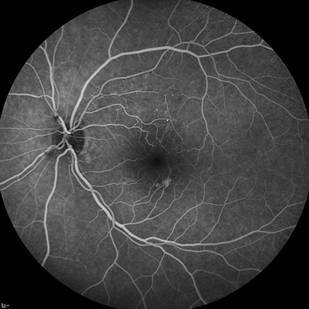
Fluorescein angiography is a diagnostic procedure (similar to retinal photography) in which a special camera takes a series of photographs of the light-sensitive tissues at the back of the eye.
As its name suggests, the procedure involves the use of a special dye called sodium fluorescein, which is injected into the patient and travels through the bloodstream to the circulation of the eye’s fundus. The blood mixed with fluorescein is then stimulated, producing a strong light emission that allows the circulation of the retinal and choroidal vessels to be recorded photographically.
Fluorescein angiography detects abnormalities in blood vessels, structural defects in vessel walls, the appearance of new vessels, and abnormal sites of leakage or inflammation.